ElectroPhysiology Patch Clamp Setup Overview
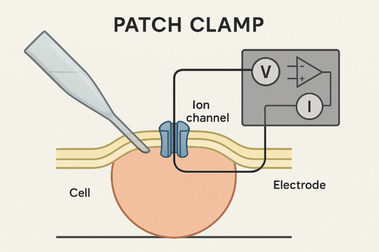
A patch clamp setup is a sophisticated ElectroPhysiology rig used to study the electrical properties of biological membranes, particularly the activity of ion channels. The technique, developed by Erwin Neher and Bert Sakmann (who were awarded the Nobel Prize for their work), allows for the recording of tiny electrical currents (in the picoampere range) that flow through single or multiple ion channels.
Key Components of a Patch Clamp Setup
A typical patch clamp rig includes several essential components working in concert to create a stable and low-noise environment for sensitive measurements.
Data Acquisition System (Digitizer & Software): The amplified analog signal from the patch clamp amplifier is converted into a digital signal by a digitizer. Specialized software then records, displays, and analyzes the data. Example acquisition systems include Scientific Solutions’ LabMaster DMA, LabMaster PRO and the LabMaster EP.
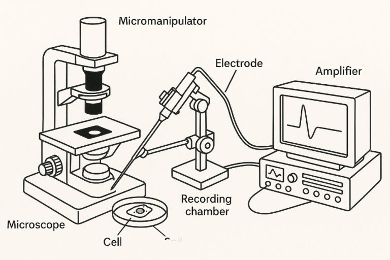
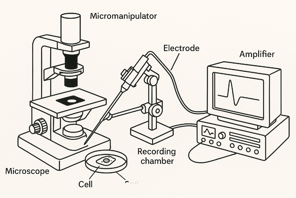
Microscope: A high-quality microscope is crucial for visualizing the cells or tissue slices to be recorded. Inverted or upright microscopes with specialized optics (e.g., DIC or oblique illumination) are often used to clearly identify and target individual cells.
Micromanipulator: This is a precision device that allows for the fine-tuned movement of the patch pipette. The manipulator must be incredibly stable and free from drift to position the pipette accurately against a cell membrane and maintain a “giga-seal.”
Patch Pipette (Micropipette): This is a hollow glass tube that has been pulled to a very fine tip, just a few micrometers in diameter. It is filled with an electrolyte solution and contains an electrode. The pipette is used to make a tight seal with the cell membrane.
Patch Clamp Amplifier: This is the heart of the electrical recording system. It consists of a headstage and a main amplifier. The headstage is mounted close to the pipette and cell, minimizing noise and amplifying the weak electrical signals. The main amplifier processes these signals, allowing for voltage or current clamping.
Electrodes:
Recording Electrode: An Ag/AgCl electrode is immersed in the electrolyte solution within the patch pipette.
Reference (Ground) Electrode: A second electrode is placed in the bath solution surrounding the cell. This completes the electrical circuit.
Perfusion System: This system is used to continuously flow fresh solution over the cells or tissue, which is essential for maintaining cell viability and for applying drugs or other compounds to the cell.
Anti-Vibration Table and Faraday Cage: Because the electrical signals being measured are so small, the entire setup must be isolated from environmental noise. An anti-vibration table dampens mechanical vibrations, and a Faraday cage shields the rig from electromagnetic interference, such as from power lines and other electronic equipment.